Retinal Tears & Detachments
The retina is a thin layer of light-sensitive nerve fibers and cells that covers the inside and back of the eyeball.
Description
The retina is a thin layer of light-sensitive nerve fibers and cells that covers the inside and back of the eyeball. For us to see, light must pass through the lens of the eye and focus on the retina. The retina then acts like a camera, taking a picture and transmitting the image through the optic nerve to the brain.
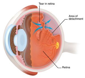
The vitreous fluid, the gel-like material that fills the eyeball, is attached to the retina around the back of the eye. If the vitreous changes shape, it may pull a piece of the retina with it, leaving a retinal tear. Once a retinal tear occurs, vitreous fluid may seep between the retina and the back wall of the eye, causing the retina to pull away and resulting in a retinal detachment.

Symptoms
The signs of a serious retinal tear or detachment can include a blind spot in your vision, blurred vision, shadowy lines, flashes of light, and floaters (spots).
Treatment
If a tear or a hole is treated before detachment develops or if a retinal detachment is treated before the central part of the retina (macula) detaches, you’ll probably retain much of your vision. Therefore, preventative care and early detection techniques are key to good maintenance of eye health.
However, if retinal holes, tearing, or detachment is advanced or serious, surgery is the only effective therapy and treatment should be sought immediately. Dr. Fox can tell you about the various risks and benefits of your treatment options. Together we can determine what treatment is best for you.

